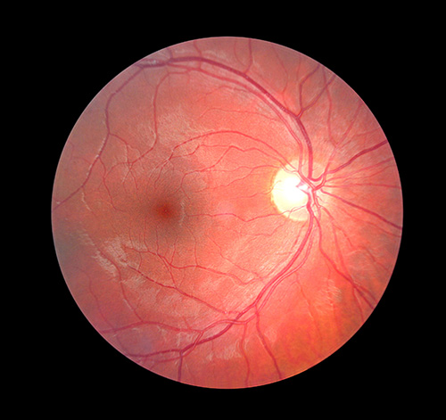
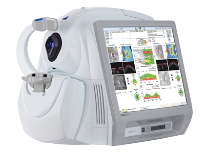
Retinal Imaging
In an effort to bring the highest level of eye care to our patients Dr. Weber uses Retinal Photography and Optical Coherence Tomography to diagnose, document and manage retinal disease.
Many eye problems can develop without warning and progress with no symptoms. Early on, you might not notice any change in your vision. However, diseases such as macular degeneration, glaucoma, retinal tears or detachments, as well as other health problems such as diabetes and high blood pressure, can be detected with a dilated examination of the retina. The retina is the part of your eye that catches the image of what you are looking at, similar to the film in a camera.
Retinal imaging provides:
- A scan to confirm a healthy eye or detect the presence of disease.
- An overview or map of the retina, giving your eye doctor a more detailed view than he can achieve by other means.
- The opportunity for you to view and discuss the images of your eye with your doctor at the time of your exam.
- A permanent record for your medical file, enabling the doctor to monitor changes in your condition on sequential examinations.
Retinal imaging includes: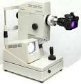
Fundus Photography - digital photo documentation of the retina
OCT - Optical Coherence Tomography, is a LASER scan of the retina which produces a 3-dimensional image primarily used in the management of glaucoma and diseases of the macula.
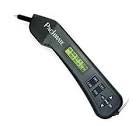
Autorefractor
An Autorefractor or automated refractor is a computer-controlled instrument used during an eye examination to provide an objective measurement of a person's refractive error (ie: prescription for glasses). This is achieved by measuring how light is changed as it enters a person's eye. This technology also enables the doctor to access the refractive status of non verbal patients such as young children and stroke victims.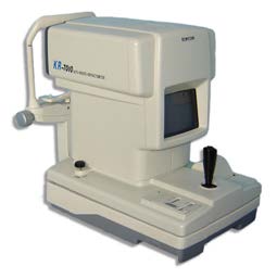
Visual Field Analyzer
The automated Visual Field Analyzer is a subjective test, which maps an individual’s field of vision. It is used extensively in the diagnosis and management of glaucoma, macular degeneration, side effects of certain medications as well as many neurological conditions such as stroke and brain tumors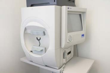
Pachymeter
A pachymeter is an ultrasound device which measures the thickness of an individuals cornea. Primarily used in the diagnosis of Glaucoma